Imagine finding out that the lining of your esophagus has changed. It sounds scary, but for millions of people, this is a manageable reality known as Barrett's esophagus, a condition where chronic acid reflux causes the normal tissue in the lower esophagus to be replaced by tissue similar to what lines the intestine. While it is a precancerous state, it is not cancer itself. The real concern lies in its potential to progress to esophageal adenocarcinoma, a serious type of cancer. However, with modern surveillance and treatment options like ablation, you can significantly reduce this risk. This guide breaks down what dysplasia means, who is at risk, and how procedures like radiofrequency ablation work to protect your health.
What Is Barrett’s Esophagus and Why Does It Matter?
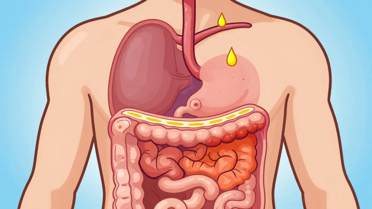
Barrett's esophagus develops when you have long-standing gastroesophageal reflux disease (GERD). Over time, the constant exposure to stomach acid damages the squamous cells that line your esophagus. In response, your body replaces them with columnar cells, which are more resistant to acid but carry a higher risk of turning into cancer. According to the American Gastroenterological Association, about 5.6% of adults in the U.S. have this condition, affecting roughly 3.3 million people. Only 10-15% of people with chronic GERD develop Barrett's, so having heartburn doesn't automatically mean you have it. But if you do, understanding the next steps is crucial because early detection can improve survival rates from 20% in advanced stages to over 80%.
Who Is at Risk for Barrett’s Esophagus?
Not everyone with reflux gets Barrett's esophagus. Certain factors make you more susceptible. If you are male, over 50, white, or have a family history of the condition, your risk goes up. Obesity, particularly around the abdomen, increases pressure on the stomach, pushing acid upward. Smoking is another major risk factor. Interestingly, alcohol does not appear to increase the risk, and some studies suggest that *Helicobacter pylori* infection might actually lower it by reducing stomach acid production. If you have weekly GERD symptoms for five years or more, your prevalence rate jumps to 3%, compared to 0.8% in the general population. Knowing these risks helps you decide if you need screening.
Understanding Dysplasia: The Warning Sign
Dysplasia refers to abnormal cell growth in the Barrett's tissue. It is graded based on how close the cells are to becoming cancer. There are three main stages:
- No dysplasia: The cells look abnormal due to metaplasia but are not pre-cancerous. The annual risk of progressing to cancer is low, around 0.2-0.5%.
- Low-grade dysplasia (LGD): Some cells show early signs of change. This increases the risk to 6-19% per year. Diagnosis here can be tricky, as pathologists sometimes disagree on whether changes are truly dysplastic or just inflammation.
- High-grade dysplasia (HGD): The cells are very abnormal and close to becoming invasive cancer. The risk of progression skyrockets to 23-40% per year without treatment.
Long-segment Barrett's esophagus (more than 3cm) also carries a higher risk. A study found that segments longer than 10cm increase the risk of progression by over 10 times compared to shorter segments. Persistent acid exposure despite medication further raises this danger.
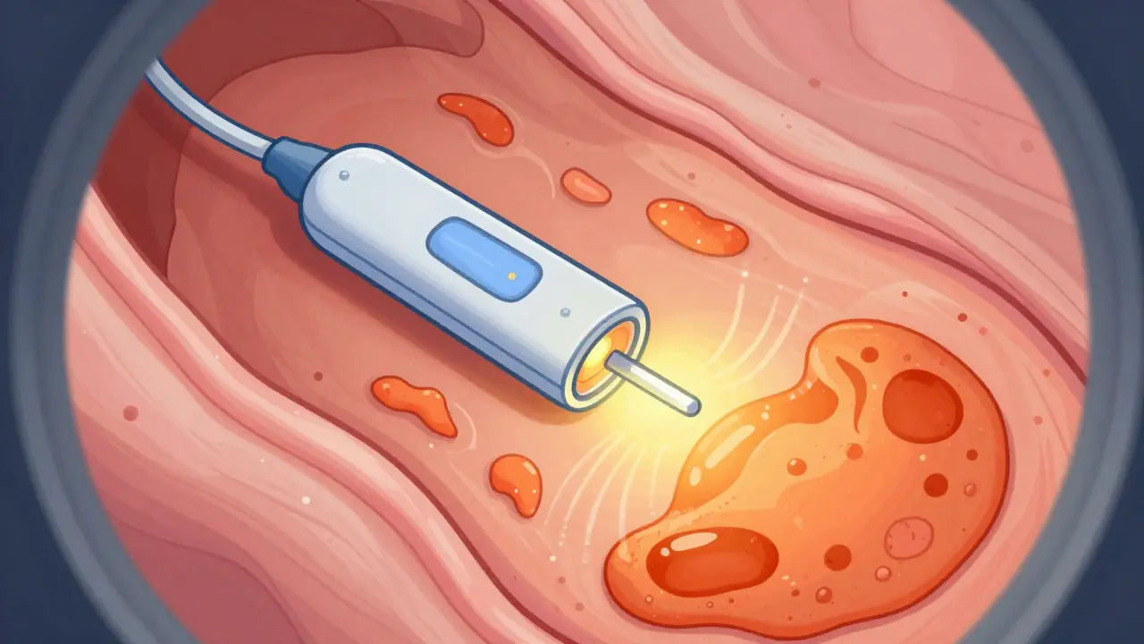
Ablation Options: Treating Dysplastic Tissue
If you have confirmed dysplasia, especially high-grade, doctors usually recommend endoscopic eradication therapy rather than just watching and waiting. These procedures destroy the abnormal tissue, allowing healthy cells to grow back. Here are the primary options:
| Method | How It Works | Efficacy (Dysplasia Eradication) | Key Risks/Side Effects |
|---|---|---|---|
| Radiofrequency Ablation (RFA) | Uses heat energy via a catheter (HALO system) to destroy tissue. | ~91.5% complete eradication of intestinal metaplasia; 87.9% for dysplasia. | Strictures (6.2%), chest pain, rare bleeding. |
| Cryoablation | Freezes tissue using nitrous oxide (-85°C) via a balloon catheter. | ~82% complete eradication of dysplasia. | Lower stricture rate (2.8%), good for prior strictures. |
| Photodynamic Therapy (PDT) | Light-sensitive drug + laser light destroys cells. | ~77% dysplasia eradication. | High stricture rate (17%), skin photosensitivity (15-20%). |
| Endoscopic Mucosal Resection (EMR) | Surgically removes visible lesions during endoscopy. | 93% en bloc resection for small lesions (<2cm). | Bleeding (5-10%), perforation (2%). |
RFA is currently the gold standard. It uses controlled heat to ablate the surface layer. Most patients need multiple sessions. Cryoablation is gaining traction, especially for patients who already have strictures, as it has a lower risk of causing new ones. PDT is less common now due to side effects like needing to avoid sunlight for weeks. EMR is often used first to remove any visible bumps before ablating the flat surrounding tissue.
Success Rates and Patient Experiences
How well do these treatments work? Data shows that RFA reduces the risk of progression to adenocarcinoma by 90% compared to surveillance alone. In clinical trials, over 77% of patients achieved complete eradication of intestinal metaplasia after one year. However, success isn't guaranteed for everyone. Recurrence can happen, which is why ongoing surveillance is necessary even after successful ablation. Combining RFA with high-dose proton pump inhibitors (PPIs) has been shown to reduce recurrence risk to 8.3% at three years, compared to 24.7% with standard doses.
Patient experiences vary. Many report relief from chronic reflux symptoms like coughing after treatment. Others face challenges with strictures-narrowing of the esophagus-which may require dilation procedures. One patient shared that while they achieved complete eradication after three RFA sessions, they needed four dilations for strictures, describing the chest pain as worse than their original symptoms. This highlights the importance of discussing potential complications with your doctor beforehand.

Costs and Accessibility
The cost of ablation can be significant. An RFA procedure averages $12,450, including equipment and physician fees, while cryoablation costs around $9,850. Insurance coverage varies, but Medicare bundles EGD with ablation at $1,850. Despite the upfront cost, RFA is considered cost-effective over five years due to lower retreatment rates. Access is another issue. While 85% of academic centers offer ablation, only 42% of rural community practices do. This gap means patients in underserved areas may face longer wait times or travel burdens, contributing to higher mortality rates in those regions.
Future Directions and New Technologies
The field is evolving rapidly. New devices like the Barrx iCAP cryoablation system offer real-time temperature monitoring for better precision. Artificial intelligence is being integrated into endoscopy to help detect dysplasia with 94% accuracy, far surpassing human eye performance in some studies. Molecular biomarkers, such as TFF3 methylation testing, could soon help identify high-risk patients more accurately, potentially reducing unnecessary procedures by 30%. These advancements promise safer, more effective care for those living with Barrett's esophagus.
Does everyone with Barrett's esophagus need ablation?
No. Ablation is typically recommended for patients with confirmed low-grade or high-grade dysplasia. For non-dysplastic Barrett's, regular surveillance endoscopies are usually sufficient because the annual risk of cancer progression is very low (0.2-0.5%). Doctors weigh the benefits of preventing cancer against the risks and costs of the procedure.
What are the most common side effects of radiofrequency ablation?
The most common side effect is chest pain or discomfort after the procedure. Strictures, or narrowing of the esophagus, occur in about 6% of cases and may require dilation. Bleeding and perforation are rare but serious risks. Most side effects are temporary and manageable with medication.
How many sessions of ablation do I need?
Most patients require two to three sessions of RFA, spaced several weeks apart, to achieve complete eradication. Some may need more depending on the length of the Barrett's segment and how well the tissue responds. Follow-up biopsies determine if additional treatment is necessary.
Can Barrett's esophagus come back after ablation?
Yes, recurrence is possible. Even after successful eradication, the underlying tendency for metaplasia remains. This is why lifelong surveillance endoscopies are recommended. Taking high-dose PPIs can help reduce the risk of recurrence, keeping it as low as 8.3% at three years in some studies.
Is cryoablation better than RFA?
Cryoablation is not necessarily "better" but may be preferred in specific cases. It has a lower rate of stricture formation, making it ideal for patients who already have strictures or cannot tolerate them. However, RFA generally has higher efficacy rates for complete eradication. Your gastroenterologist will choose the best option based on your individual anatomy and medical history.